Pneumoperitoneum caused by tuberculosis
DOI:
https://doi.org/10.31837/cir.urug.5.2.1Keywords:
pneumoperitoneum, tuberculosis, surgeryAbstract
86-year-old male who came to the emergency room due to lower limb edema. On examination, he presented discreet abdominal discomfort, with no other accompanying symptoms.
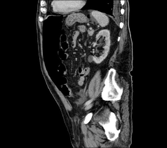
The analysis shows a hemoglobin of 8.2 g / dL with a hematocrit of 24%. A chest and abdominal CT scan was performed (Fig. 1) that revealed bilateral pulmonary nodules with an inflammatory appearance, pleural effusion and massive pneumoperitoneum, without evidence of rupture of the hollow viscus. He is kept on an absolute diet and antibiotic therapy and parenteral nutrition are started.
Cultures are extracted, mycobacteria appearing in the bronchoalveolar lavage. He is diagnosed with disseminated tuberculosis. The antibiotic treatment is adjusted and the patient improves progressively, being discharged from hospital 10 days after the onset of the symptoms. In the follow-up CT scan at 3 months, the pneumoperitoneum has disappeared.
The most frequent origin of spontaneous non-surgical pneumoperitoneum is the thorax (due to tuberculosis, mechanical ventilation, barotrauma, pulmonary contusion, chronic obstructive pulmonary disease…), there are also abdominal causes such as intestinal cystic pneumatosis. Between 5 and 14% of patients with spontaneous pneumoperitoneum can be managed conservatively, without surgery. Some series show that no visceral perforation is evident in up to 44% of non-surgical pneumoperitoneums. It is therefore a cause of non-surgical pneumoperitoneum that, with an adequate clinical and therapeutic approach, makes it possible to avoid surgical intervention. 1,2,3,4,5
Downloads
Metrics
References
2. Martínez-Hernández-Magro P, Lazarini-Díaz-Barriga JA, Mendoza-Suárez J, Lemus-Sánchez G. Neumoperitoneo no quirúrgico. Anales de Radiología México. 2019;18:207-211. DOI: 10.24875/ARM.19000106
3. Larrañaga I, Meneu JC, Díaz G, Mendía E, Rey A, Fresneda V. Neumoperitoneo no quirúrgico. Cir Esp. 2000;67:5:411-523. En: https://www.elsevier.es/es-revista-cirugia-espanola-36-articulo-neumoperitoneo-no-quirurgico-10573 DOI: 10.31837/cir.urug/5.2.1 Cir. Urug. Vol. 5 No. 2 Jul. – Dic. 2021 1-3
4. Ahmad QA, Sarwar MZ, Fatimah N, Ahmed AS, Changaizi SH, Ayyaz M. Acute Presentation and Management of Abdominal Tuberculosis. J Coll Physicians Surg Pak. 2020 Feb;30(2):129-133. DOI: 10.29271/jcpsp.2020.02.129
5. Kentley J, Ooi JL, Potter J, Tiberi S, O'Shaughnessy T, Langmead L et al. Intestinal tuberculosis: a diagnostic challenge. Trop Med Int Health. 2017 Aug;22(8):994-999. DOI: 10.1111/tmi.12908
Published
How to Cite
Issue
Section
License

This work is licensed under a Creative Commons Attribution-NonCommercial 4.0 International License.
All articles, videos and images published in Revista Cirugía del Uruguay are under the Creative Commons CC licenses, which is a complement to the traditional copyright, in the following terms: first, the authorship of the referred document must always be acknowledged and secondly none of the article or work published in the journal may have commercial purposes of any nature. The authors retain their copyrights and give the magazine the right of first publication of their work, which will be simultaneously subject to the Creative Commons Attribution-NonCommercial 4.0 International License license that allows the work to be shared whenever the initial publication is indicated in this journal.